1.2. History
1.2.1. Learning Objectives
Upon completion of this section, the student will be able to:
describe landmark events that launched the field of CAS
1.2.2. Discussion
A review paper by Terry Peters and Kevin Cleary gives a good introduction to the field of Image-guided Interventions [PetersCleary2010].
In this section here, we just cover a few key events, before delving more deeply into a selection of System Examples for a more detailed study, of what makes up a CAS system.
1.2.3. Pre X-ray Guidance
Before the advent of interventional imaging the only physical way to determine what was inside the human body was to cut it open and use your eyes / hands. Thus surgery is both the diagnostic and theraputic tool.
1.2.4. First X-ray Guidance
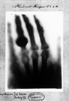
8th November 1895 - Röntgen documented the first X-ray experiment. The experiment was first published on 28th Dec 1895 (Wikipedia). The first version that we can actually find, in English, was published on 14th Feb 1896 [Roentgen1896].

Fig. 1.4 The first medical X-ray by Wilhelm Röntgen of his wife Anna Bertha Ludwig’s hand, Wilhelm Röntgen. [Public domain], from Wikimedia.
11th Jan 1896 - the first clinical use of X-rays by John Hall-Edwards, Birmingham, UK, to remove a needle from a hand (Wikipedia).
7th February 1896 - the first surgical use of X-rays by Professor John Cox, Montreal, Canada, where X-rays were used to remove a bullet from a patients leg [Cox1896].
1.2.5. Stereotactic Surgery
1908 - Horsley and Clarke described the stereotactic frame [HorsleyClarke1908], defining anatomical coordinates, leading to stereotactic surgery. “Stereo” - meaning solid, and “taxis” meaning ordered.

Fig. 1.5 Stereotactic apparatus used by Sir Victor Horsley and Richard Clarke. Made by Swift and Son, London, c1905, from sciencemuseumgroup.org.uk, licensed under CC BY-NC 4.0.
This led to other frames, using for example spherical coordinates:

Fig. 1.6 Figure Arc for Leksell Stereotactic System, c1997. Frame for Leksell Stereotactic System, c1997. from sciencemuseumgroup.org.uk, licensed under CC BY-NC 4.0.
and with the advent of CT imaging in the 1970’s, to frames that could be imaged, to more easily map from image coordinates to physical coordinates. This means, you can understand where your physical tools are in relation to pre-operative imaging, or vice-versa.

Fig. 1.7 Stereotactic frame with N-localisers, by Kirigiri, on wikimedia, licensed under CC BY-SA 3.0.
This is an example on YouTube, from the Institute for Cancer Genetics and Informatics channel, where stereotactic coordinates are used for brain biopsy.
1.2.6. Frameless Stereotaxy
So, the advent of CT scanning in the 1970s and the modern PC in the 1980s led to the concept of frameless stereotaxy [PetersCleary2010], first in the operating microscope [Roberts1986] and then with a mechanical arm for a tracker, with the display using the a 4-quadrant view [Galloway1993].
1.2.7. Surgical Planning
Pioneered by Terry Peters et al. [Peters1987], [Peters1989]. The aim here was to integrate multi-modality imaging (MR/CT/DSA) via fiducials visible in each image, and also to provide a stereoscopic display for better visualisation of DSA.